Limb Lengthening &
Deformity Corrections
Expert Surgical Solutions for Complications
Pediatrics & Adults

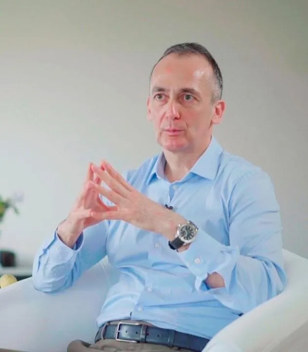
About Dr. Metin Küçükkaya
Dr. Metin Küçükkaya is a globally recognized orthopedic surgeon specializing in deformity correction, limb lengthening, and orthopedic trauma.
Over the past 30 years, he has not only witnessed but led the evolution of surgical techniques —advancing from classical Ilizarov methods to the most sophisticated intramedullary lengthening nails, plates, and systems for both adult and pediatric patients.
His specialties include:
Primary orthopedic deformities and limb shortening in Adults & Pediatrics.
Orthopedic trauma and its complications, including malunions, nonunions, bone infections, and limb length discrepancies—particularly those resulting from inadequate treatment by other healthcare providers in both Adult & Pediatric cases.
There’s no such thing as an "impossible case"
"Only one that wasn’t planned well"
A Personalized,
Hands-on Approach to Patient Care
Under the Guidance of Dr. Metin Küçükkaya
- Custom Treatment Plans Based on Scientific Evaluation
- Meticulous Surgical Techniques Adapted to Each Patient
- Comprehensive Postoperative Support for Optimal Recovery
- An Expert Team with Over 20 Years of Specialized Experience in Limb Lengthening and Complex Deformity Correction
Office & Radiology Department
Personalized Radiological Evaluation with
"EOS Technology"
EOS technology has long been the standard for all long-standing X-rays, and Dr. Küçükkaya personally reviews and analyzes these images using advanced computer programs. Each patient receives a detailed radiological and clinical report prepared directly by Dr. Küçükkaya, ensuring a comprehensive and individualized evaluation.
Hospitals
World-Class Surgical Centers
- Procedures at Florence Nightingale and Acıbadem Maslak Hospitals
- International standards in technology, safety, and patient-centered care
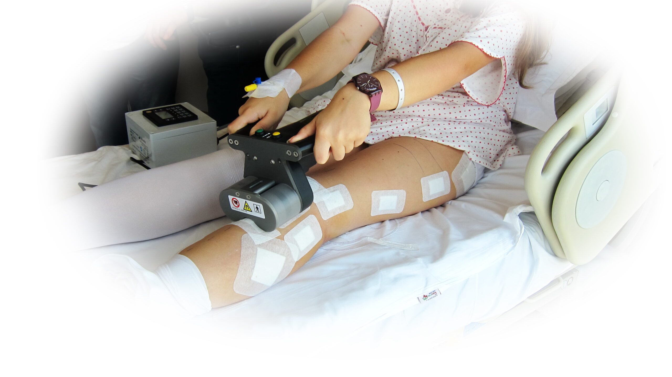
Physiotherapy & Rehabilitation
Tailored Recovery Programs
- Personalized rehabilitation plans after surgery
- Focused on each patient's specific needs for optimal recovery
Case of the month
From Cosmetic Concern to Functional Correction
Surgical Management of Mild Genu Varum
in a Young Adult
Overview:
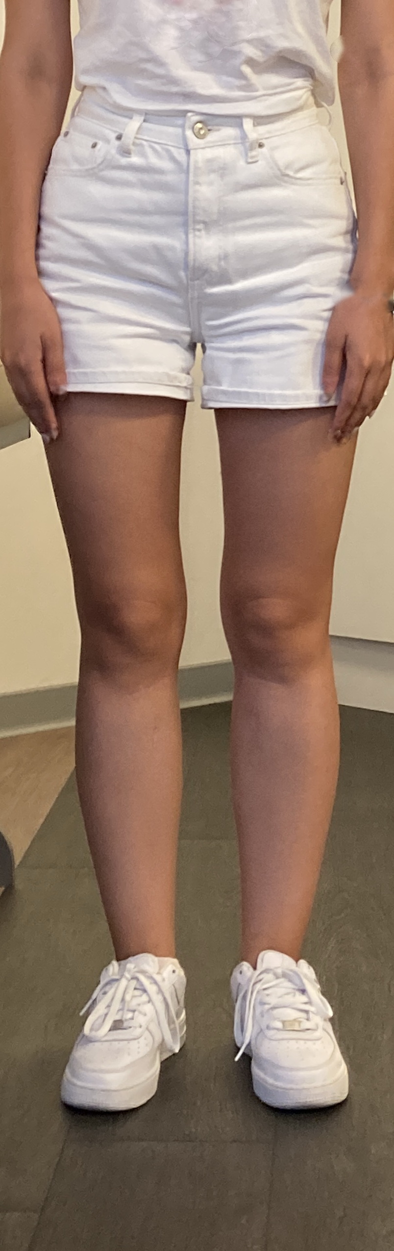
A 35-year-old patient presented with bilateral mild genu varum deformity, motivated primarily by cosmetic concerns. Although asymptomatic, the patient had a strong family history of early knee osteoarthritis, which raised concern for future joint degeneration. This case demonstrates the ethical decision-making, precise radiological planning, and scientific surgical strategy required when treating cosmetic deformity in young individuals.
🔍 Initial Clinical Findings:
- Mild bilateral tibial bowing
- No pain, instability, or activity limitation
- Family history of osteoarthritis
- Desire for improved cosmetic alignment
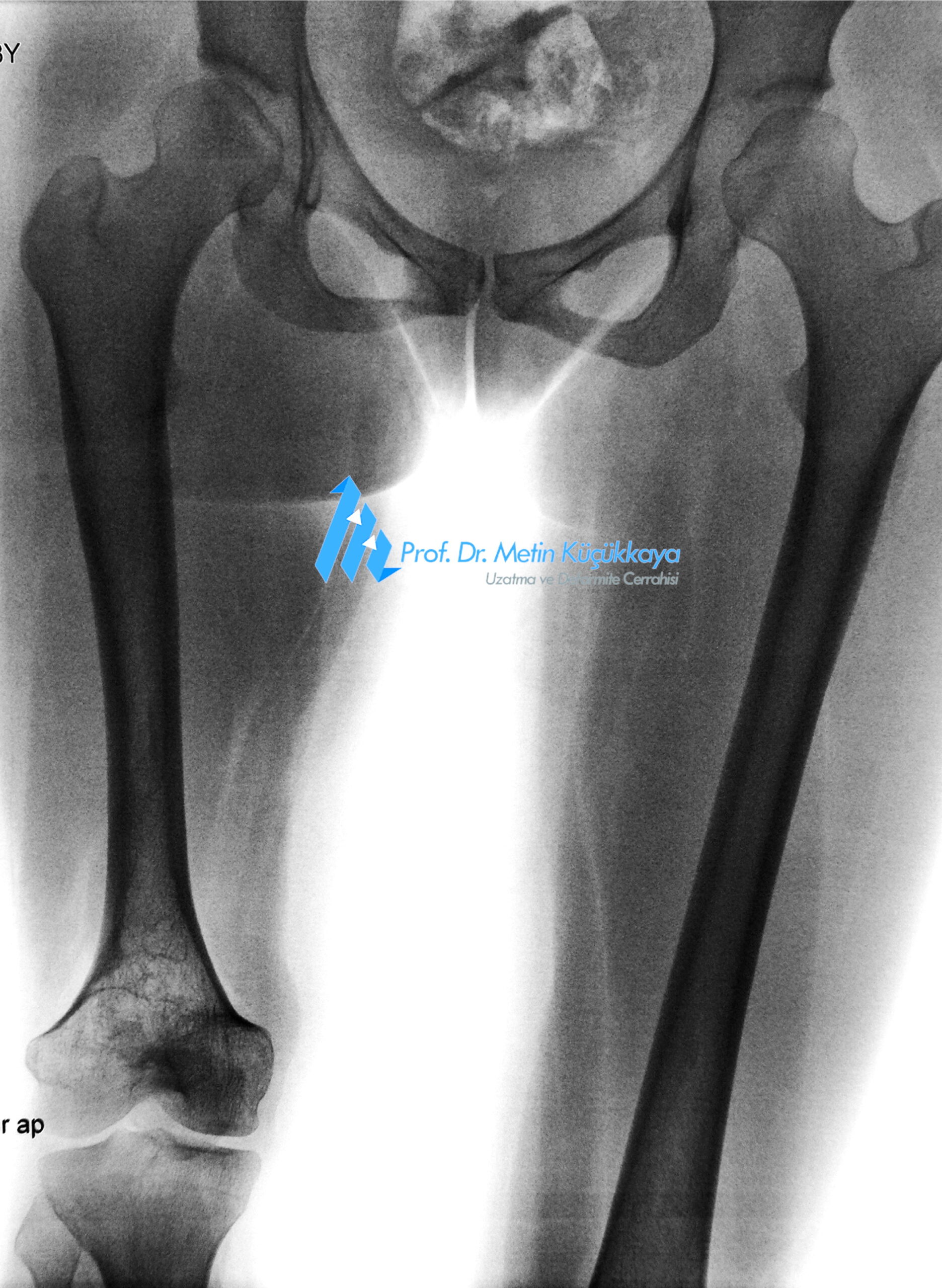
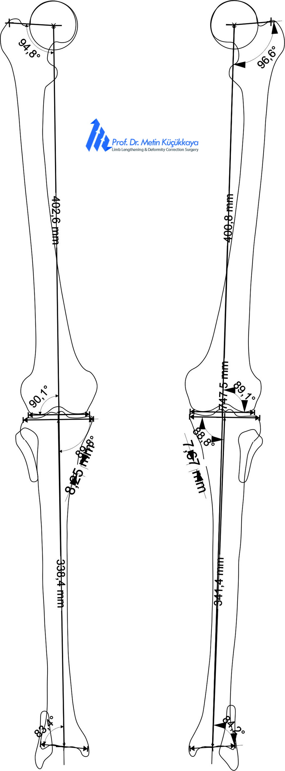
- Mechanical axis deviation confirmed on xray imaging
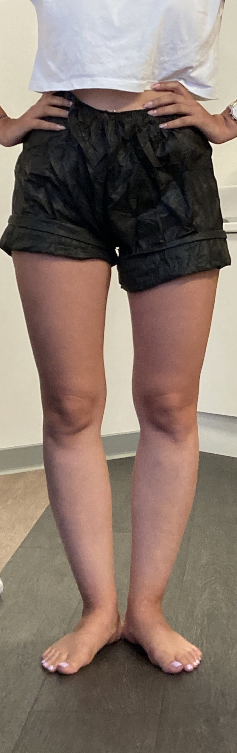
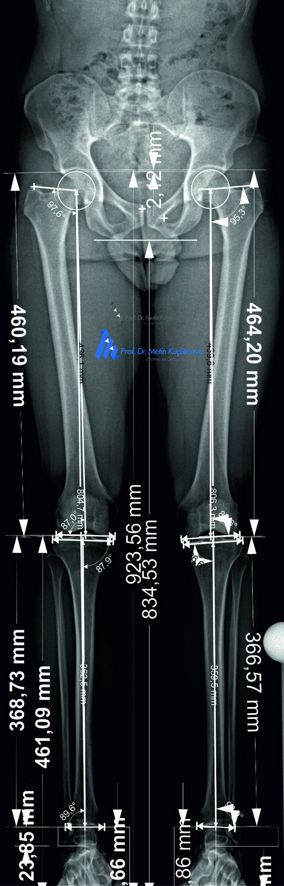
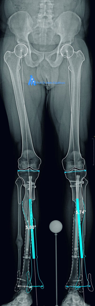
Preoperative clinical photograph and calibrated long-standing radiographs with CorelDRAW®-based measurements showing mild bilateral tibial bowing and mechanical axis deviation.
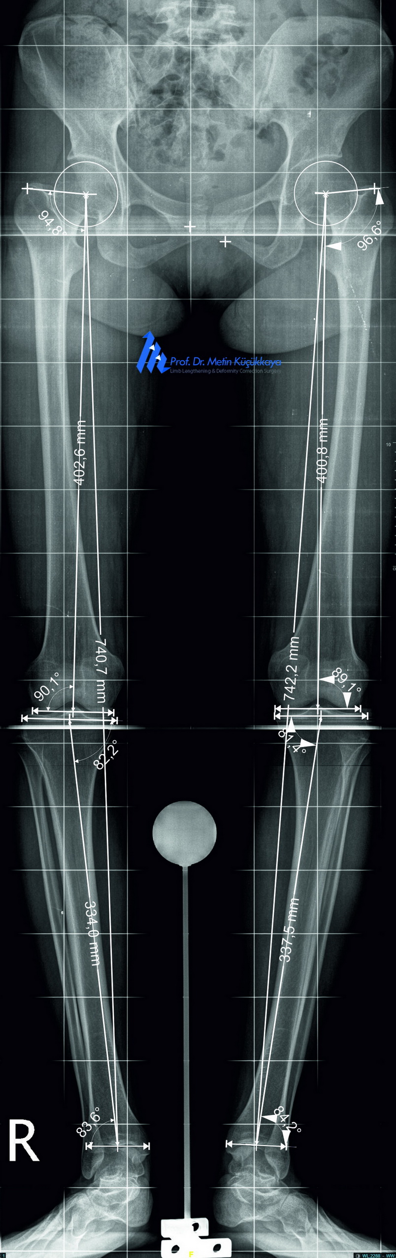
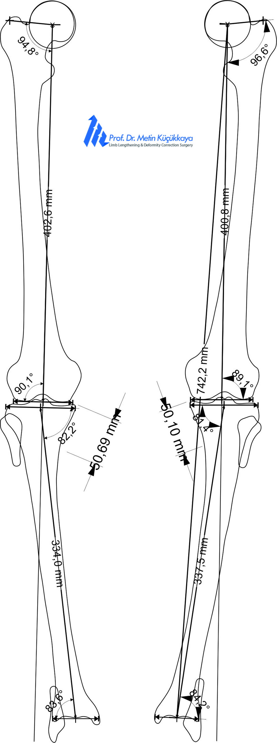
📐 Preoperative Planning:
The surgical plan was created using:
- Calibrated long-standing radiographs
- The End Point First (EPF) Method for accurate mechanical axis restoration
- CorelDRAW®–based digital preoperative planning
- Careful analysis of wedge opening, correction angle, axis deviation, and avoidance of over- or under-correction
🛠️ Surgical Treatment:
- Biplanar open-wedge high tibial osteotomy
- Opening the wedge precisely as calculated in the EPF plan
- Protection of medial cortical hinge
- Stable fixation with anatomically positioned implants
✅ Postoperative Course and Outcome:
Precise Alignment, Natural Appearance
- X-rays confirmed a precise and stable correction of the leg alignment. Healing was smooth, and the patient returned to walking and daily activities shortly after surgery. The patient reported very high satisfaction with the improved alignment and the natural cosmetic result.
💬 Clinical Insight:
- This case illustrates that cosmetic deformity correction can be ethically justified when supported by clinical risk factors such as family history of osteoarthritis. With calibrated imaging, digital planning, and precise surgical execution, we successfully transformed a cosmetic concern into a preventive and functional orthopedic correction.
Case of the month
From Complication to Correction
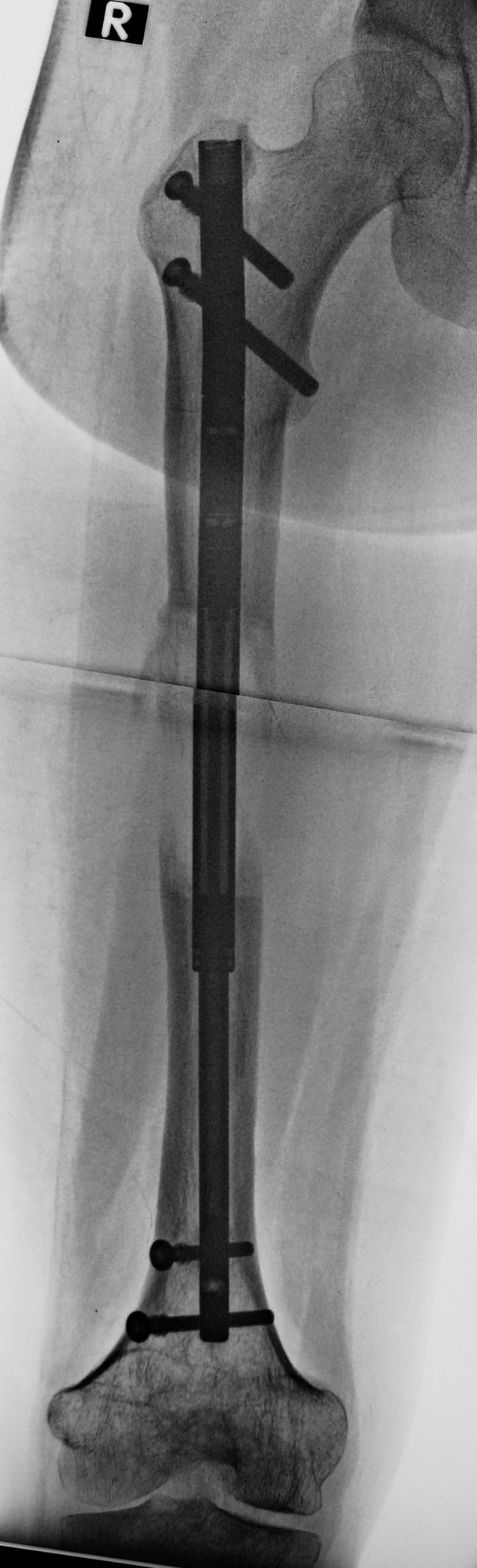
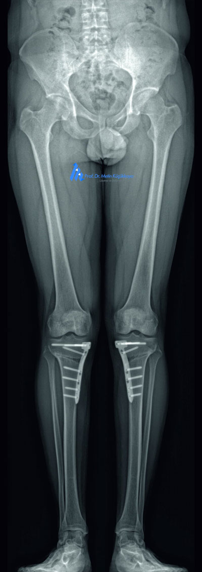
Complications After Cosmetic Tibial Lengthening
A Salvage Surgery Example
Overview:
A middle-aged patient presented with significant complications following bilateral cosmetic tibial lengthening surgery performed abroad.
🔍 Initial Clinical Findings:
- Draining wound over the anterior tibia on one side
- Contractures in both knees and ankles
- Valgus deformity in one tibia, varus in the contralateral tibia
- Bent and externally rotated intramedullary nails
- Distal migration of fibular heads and proximal migration of lateral malleoli, causing joint dysfunction
- Radiological suspicion of localized low-grade infection
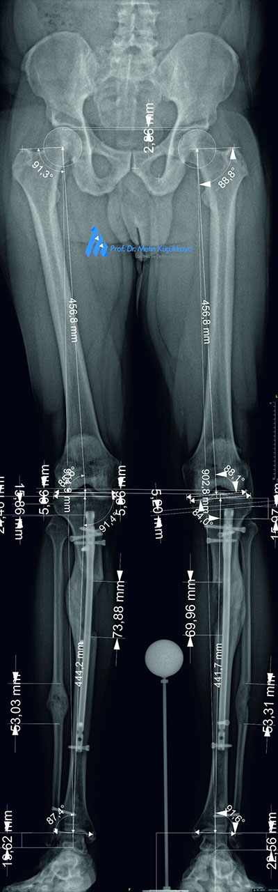
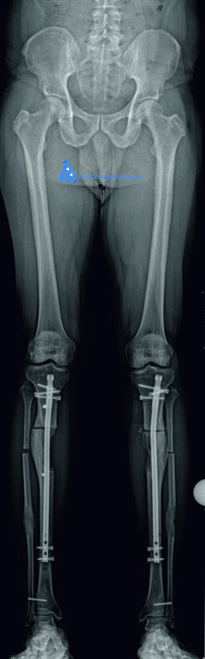
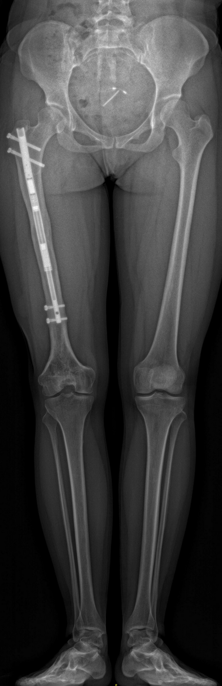
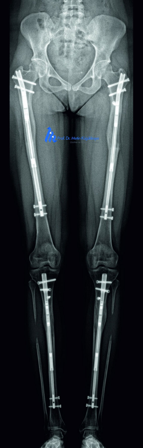
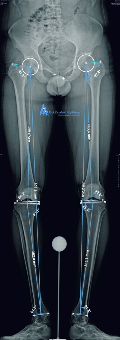
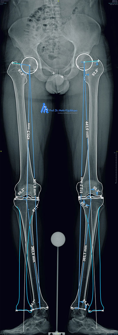
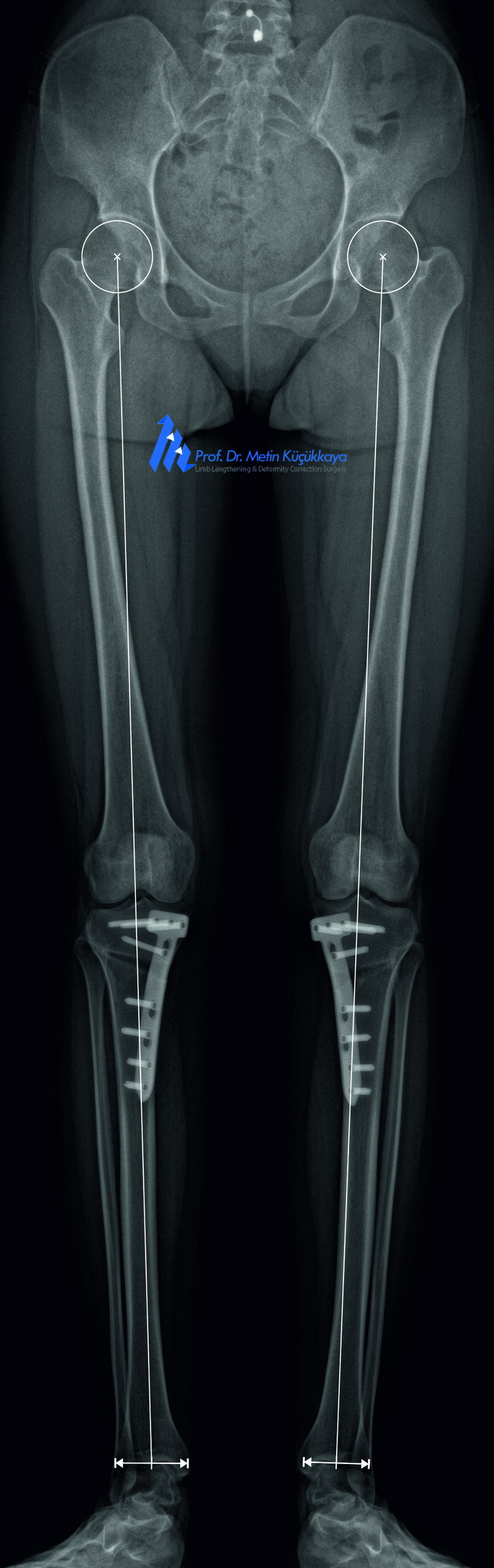
Preoperative calibrated EOS images with CorelDRAW®-based measurements (left) and planning (middle), followed by postoperative X-rays (right) showing successful correction.
📐 Preoperative Planning:
The patient underwent long-standing calibrated EOS imaging. Precice planning performed using with CorelDRAW®, mechanical axis deviation, rotation, and implant positioning were carefully analyzed. The plan involved simultaneous correction of both alignment and hardware-related complications.
🛠️ Surgical Treatment:
The surgery consisted of:
- Removal of both tibial intramedullary nails
Intraoperative debridement of the medullary canals - Sampling for culture and antibiogram
- Insertion of absorbable calcium sulfate (Stimulan®) loaded with antibiotics
- Correction of tibial deformities with pre-bent, antibiotic-coated intramedullary nails
✅ Postoperative Course and Outcome:
Radiographs confirmed anatomical correction and implant stability. Wound healing was achieved, and the patient gradually returned to independent ambulation.
💬 Clinical Insight:
"This case highlights the potential risks associated with unregulated cosmetic lengthening procedures and the critical role of expert salvage strategies in restoring both form and function. A combination of calibrated imaging, digital planning, and staged surgical execution enabled us to turn a severe complication into a successful clinical outcome."
Our Services
Choose Your Treatment
BestForLengtheningComplications
Complications After Cosmetic Limb Lengthening
Deformities & Malunion
- Angular - Torsional
Bone Formation Failure After Lengthening
Discrepancy after lengthening
Asymmetrical Bone Lengths without Discrepancy
Infection – Osteomyelitis
Neuro-vascular complications
Joint contractures
Limping
Physiological and Psychological Maladaptation After Lengthening
BestForOrthoComplications
Complications After Trauma or Infection
Nonunion - Malunion after trauma
Bone infections - Osteomyelitis
Bone defects
- Trauma
- Tumor resection
- Ostemyelitis
Joint contractures
Neuro-vascular complications
BestForCosmeticLengthening
Cosmetic Stature Lengthening
Cosmetic Height Lengthening
- Femurs (Bilateral
- Tibias (bilateral)
- Femur + Tibias (Quadrilateral)
Height Lengthening Achondroplasia
Arm Lengthening
BestForDeformityCorrection
Discrepancy & Deformities
in Adults
Bowlegs in Young Individuals
Knock Knees in Young Individuals
Bowleg Correction to Prevent Osteoarthritis
Discrepancy after total hip arthroplasty
BestForKidsOrtho
Discrepancy & Deformities
in Children
Congenital limb length deformity & discrepancies
Acquired limb length deformity & discrepancies
Achondroplasia
Arm Lengthening
Lengthening - Surgery Process
Step 01
Scheduling a Consultation Appointment
- Patients begin by scheduling a consultation appointment, either virtual or in-person.
- After submitting a request, a consultation assistant will contact the patient to organize the appointment and provide necessary guidance.
- The appointment is set up based on the patient's availability and medical history.
- Initial documentation and medical records are reviewed before the consultation.
Step 02
Consultation (Virtual/In-Person)
- Initial assessment personally conducted by Dr. Küçükkaya.
- Discussion of patient goals, expectations, and possible alternatives.
- Dr. Küçükkaya personally assesses the patient’s eligibility for the proposed treatment.
Step 03
Preoperative Planning
- Detailed medical evaluation and imaging, assessed by Dr. Küçükkaya.
- Selection of the most suitable surgical technique and development of a personalized treatment plan under his direct supervision.
Step 04
2nd In-Person Consultation (Patient Education & Consent)
- Comprehensive explanation of the procedure, potential complications, and recovery expectations, led by Dr. Küçükkaya.
- The informed consent process ensures patient understanding and autonomy by discussing potential risks, benefits, and alternative treatments in a one-on-one consultation.
- Psychological counseling and support options discussed.
- Final pre-surgical preparations, including prehabilitation recommendations.
Step 05
Surgery Day
- Hospital admission and surgical setup, personally overseen.
- Surgery performed with meticulous attention to detail.
- Post-operative monitoring and care directly managed.
- Initiation of physiotherapy to promote circulation and reduce stiffness.
Step 06
Physical Therapy & Lengthening Phase
- Gradual bone distraction using an external or internal device.
- Physiotherapy and rehabilitation tailored to each patient, continuously adjusted.
- Frequent follow-ups and direct evaluations to prevent complications and ensure steady progress.
- Patients are actively involved in their rehabilitation process, receiving close guidance at every step.
Step 07
Consolidation Phase
- Bone healing and strengthening phase, supervised closely.
- Regular X-ray evaluations personally reviewed to assess progress.
- Guidance on gradually resuming daily activities while ensuring bone stability.
- Emotional and psychological support for long-term recovery.
Step 08
Device (Nail) Removal
- Performed once the bone is fully consolidated.
- Minor surgical procedure for internal device removal, ensuring a seamless experience.
- Scar revision surgery, if necessary, carried out with precision.
Step 09
Recovery & Reintegration Phase
- Post-removal rehabilitation personally supervised to optimize final functional outcomes.
- Guidance on gradually resuming daily activities while ensuring bone stability.
- Emotional and psychological support for long-term recovery.
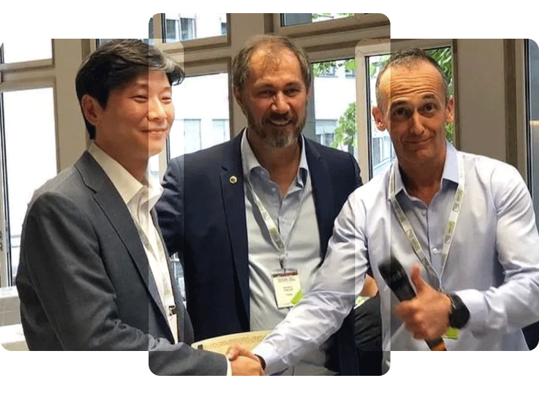
Interviews
Book a Consultation
Click Button Below to Schedule a Virtual Chat with Dr. Küçükkaya to see if LL is right for you